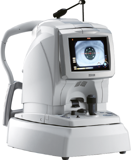
The RS-3000 Advance 2 is an OCT Spectral Domain Expert equipped with technology SLO and a function OCT-Angiography. These advanced functions make it a powerful diagnosis tool of retinal-choroidal pathologies. Thanks to an additional lens, it also makes possible analysing sections of the anterior segment.
The RS-300 Advance 2 comes with the LSO (Laser Scanning Ophthalmoscope) imaging system of the fundus, to track the retina with a high precision, thus ensuring a correct positioning of OCT and OCT-Angiography sections, considering the natural cyclotorsion of the human eye.
This technology makes also possible benefitting from an efficient HD mode, up to 120 scans that, when combined with a maximum sensibility (Ultra-Fine), provides a very accurate image from the vitreous to the choroid, whatever in retina mode or choroid mode (EDI)
The RS-300 Advance 2 is designed to perform specialised consultations. It benefits from different OCT advanced features, such as the HD mode, the manual positioning of the acquisition scans on the area of interest, the Select mode and the Rescan, to perform an additional exam, or the patient following-up, thanks to Follow-up, available with the acquisition.
Thanks to the LSO tracking, the RS-300 Advance 2 can perform the following-up exams exactly in the same conditions, using the recognition of the patient’s retina. This ensures a reliable comparative analysis, including on a single section. Over time, up to 50 exams can be compared to monitor the evolution of the pathology and/or to control the efficiency of the cure. By using trend lines and adding manually some events, such the date of the first taking of the treatment for instance, ensuring an efficient and customisable following-up is possible.
Also, the RS-300 Advance 2 is equipped with an anterior segment adapter to provide sections of the cornea and filtration angles
The NAVIS-EX software platform can analyse all the data. This interface receives the viewers dedicated to OCT and OCT-Angiography results and controls the patient database. Different map are displayed: thicknesses, global retina thickness and thickness of the complex of ganglionic cells, these thicknesses being compared with the normative 9x9 mm database, and the thickness of the nervous fibres of the optical nerve, in 6x6 mm. Also displayed are the OCT En-Face and OCT-Angiography representation, all OCT sections and 3D reconstructions.
The AngioScan makes possible analysing the vascularisation of the retina and of the choroid. The real time tracking of the retina ensures a correct positioning of the scans, to limit motion artefacts. Images are taken, either of the macula (Macula Map), or of the nervous optical head (Disc Map). The analyses provided are then different. The segmentations are adapted to the captured area, 7 differentiated areas on the macula and 4 areas on the papilla.
Thanks to the combined OCT En-Face and OCT-Angiography display, whose depth can be adjusted, and to the B-scan section (from a drop-down menu), a direct matching can be made between the structure and the tissue vascularisation.
The results of the AngioScan also include quantitative analyses. These additional parameters are used to make a diagnosis. Density and vascular perfusion values are given for each layer, as well as the information related to the central avascular zone (CAZ), which is automatically detected.
These mentions are conforms to the French regulation and may vary depending on circumstances in each country.
Brochures
Livrets de cas cliniques
OCT Denoising
OCT-A
OCT-A et Micropérimétrie
You have a project? You want a quotation? You have questions about our products?
Feel free to ask your technical sales representative.
Reliability and
safety
Technologies and
innovations
Services and
guarantees
KEY PRODUCTS
© 2021 • NIDEK SA – All rights reserved
Galerie
Ce site est réservé aux professionnels de santé