Diagnosis and therapeutic orientation
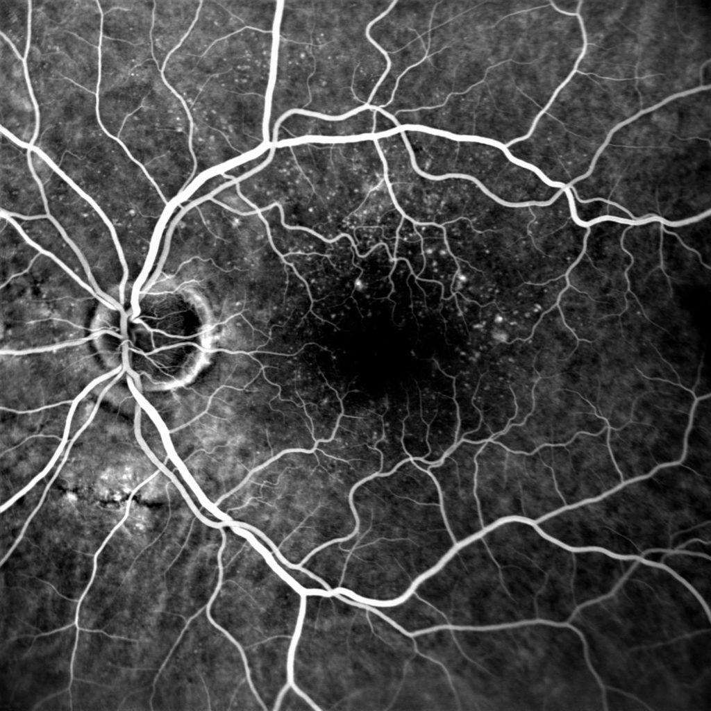
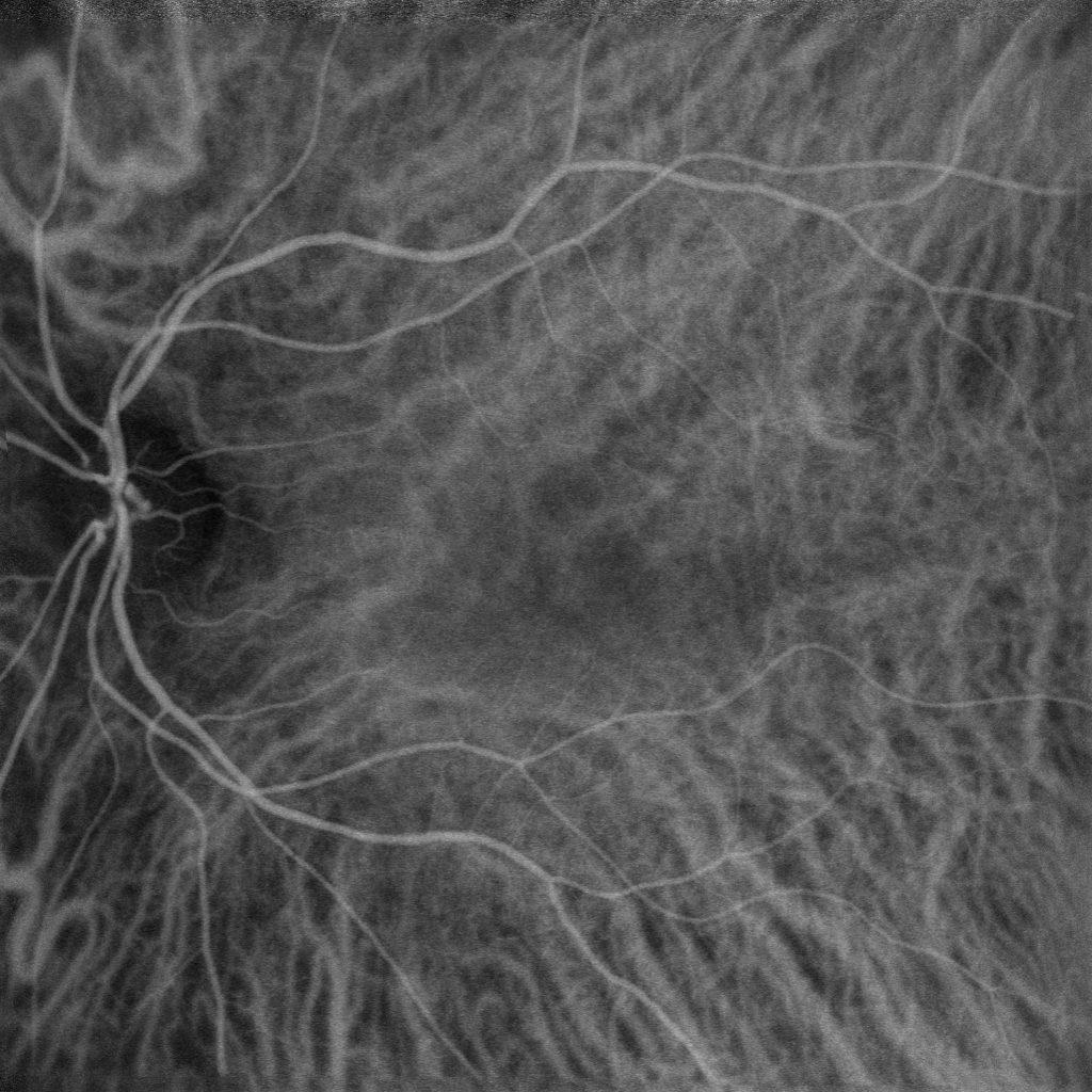
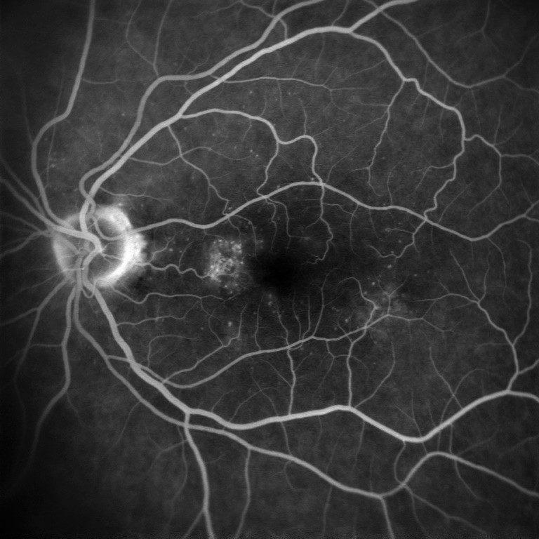
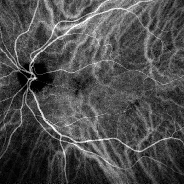
Retinal angiography is a dynamic diagnostic examination allowing the visualisation of the vascular system of the retina and detection of abnormalities that are not always visible with other fundus imaging methods.
Performed using a fluorescent contrast product (colour) injected into the patient’s blood circulation, it is therefore seen as invasive and requires a convenient equipment to perform the injection and the resuscitation in case of allergic reaction.
L’angiographie trouve son utilité pour le diagnostic et le suivi de pathologies vasculaires telles que la rétinopathie diabétique, la AMD, les uvéites postérieures…
Depending on the depth of the tissue to examine and on the pathology, two different contrast products can be used. So they define two types of angiography, the fluorescein one (FA) and the indocyanine green one (ICG).
Current angiographs, often equippedwith a confocal SLO laser system.The current angiographs, often equipped with an SLO confocal system, able to precisely focus on retina, make possible performing the fluorescein angiography only, the indocyanine green angiography only or sometimes both angiographies at the same time.
Our angiography solutions
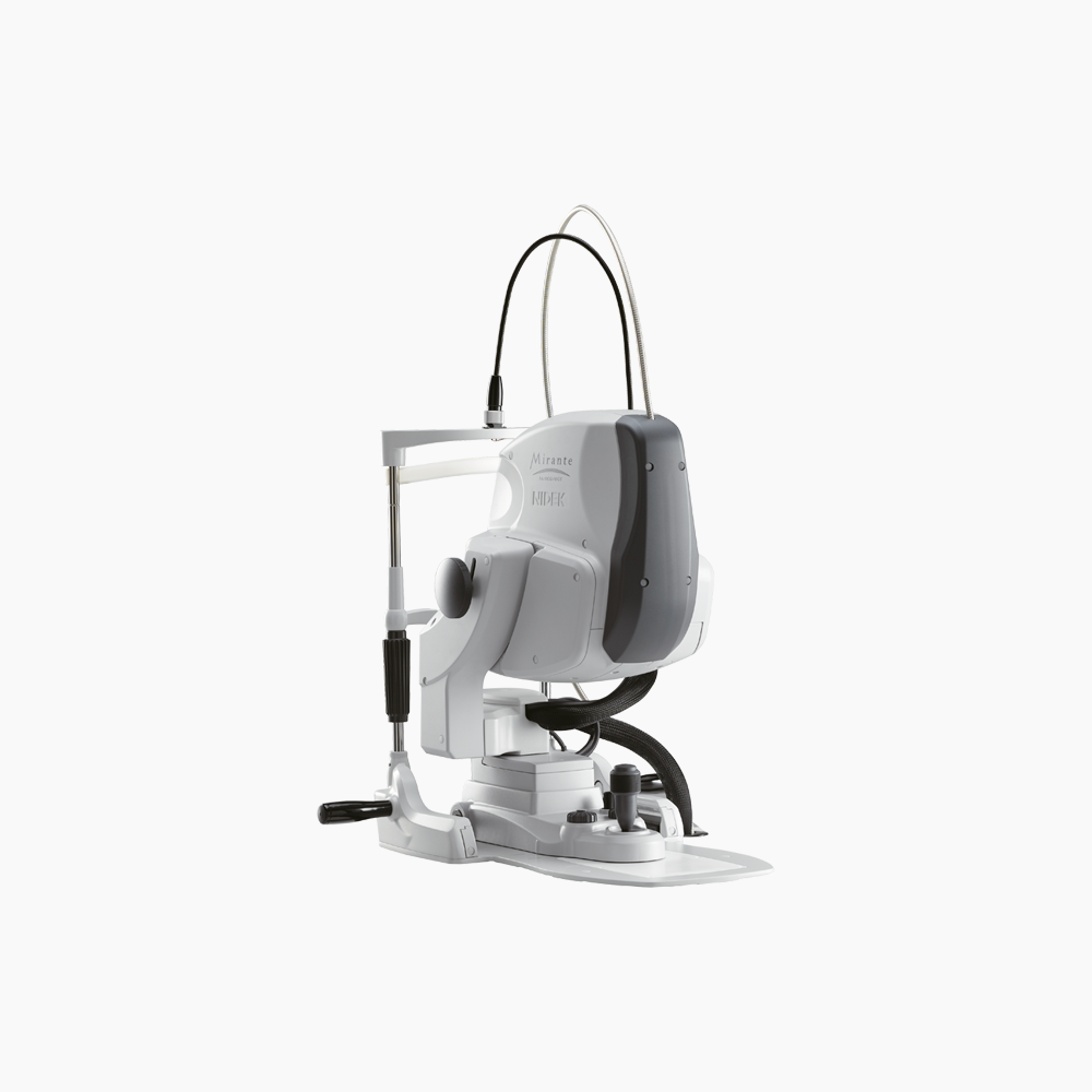
Mirante is a large-field multimodal-imaging platform designed to perform fundus imaging. It combines OCT and SLO technologies thus grouping routine imaging to detect and identify retinal-choroidal pathologies.
Mirante is a large-field multimodal-imaging platform designed to perform fundus imaging. It combines OCT and SLO technologies thus grouping routine imaging to detect and identify retinal-choroidal pathologies. Using an additional lens, performing an analysis of sections of the anterior segment is also possible.
Techniques used
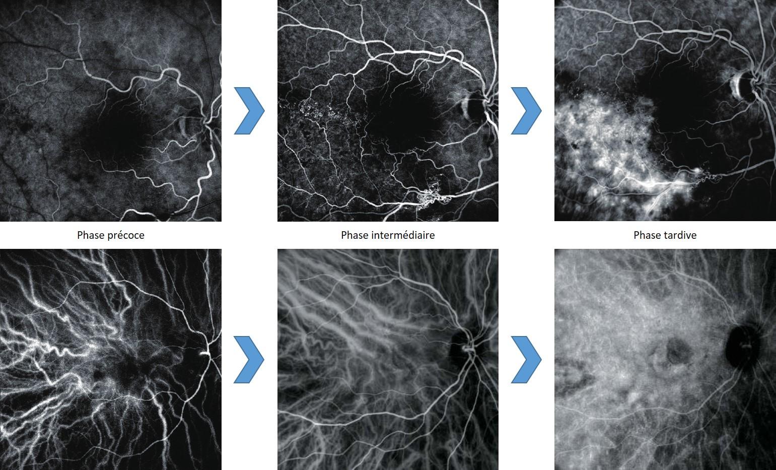
The injection angiography is the only examination making possible viewing fundus vascularisation in a dynamic way. It is then possible to analyse early, intermediate and late phases of retinal and choroidal blood circulations.
Angiography is often performed using an SLO angiograph, as this equipment can make a precise focus on retina. The purpose is detecting vascularisation anomalies, such as:
- Aneurysms
- Leaks
- Oedemas
- Ischemia
- AMD
- Diabetic retinopathy
- Vascular occlusions and tumours, etc.
The examination is performed using a contrast product injected into the patient’s blood circulation.
During the examination, the angiograph illuminates the fundus with a specific wavelength to stimulate the colour to make it visible. Angiography images then only show the colour circulation into the vascular system, thus matching the blood behaviour into the vessels.
We use angiography to make a precise diagnosis, to propose an adapted treatment and to follow-up the evolution of the patient’s pathology through a control. When a complete analysis of the structure/function of the retina is required, the angiography is always used in addition to the OCT.
This imaging technique is different from the OCT-Angiography, which is non invasive, as it derives from the OCT (scanner of the retina). This last technique remains static and for the moment its acquisition field is limited.
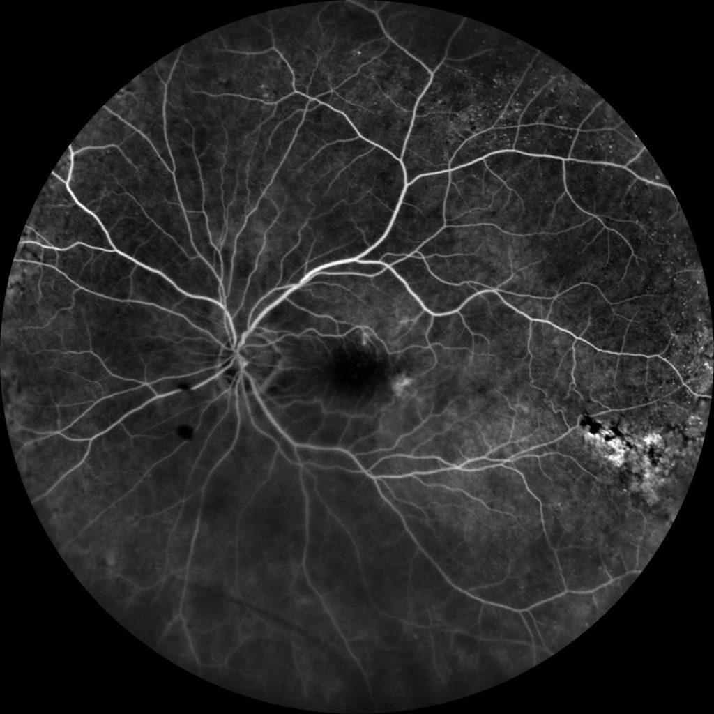
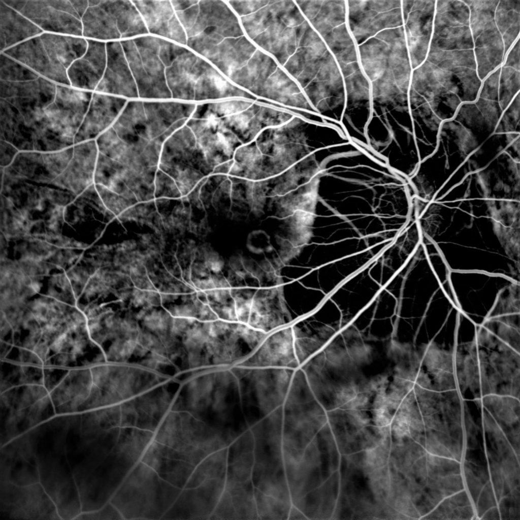
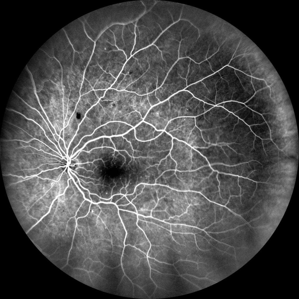
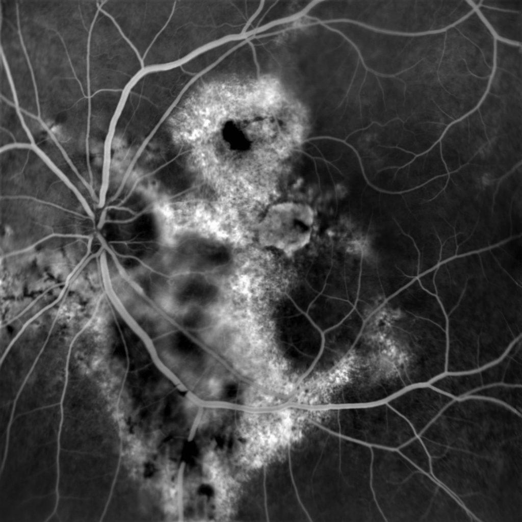
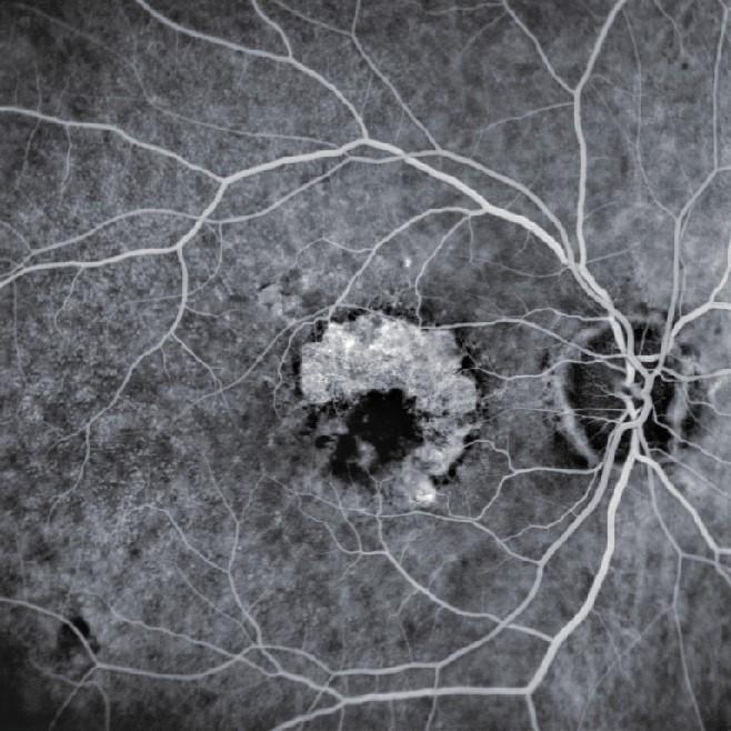
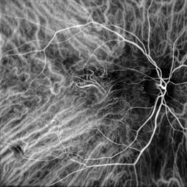
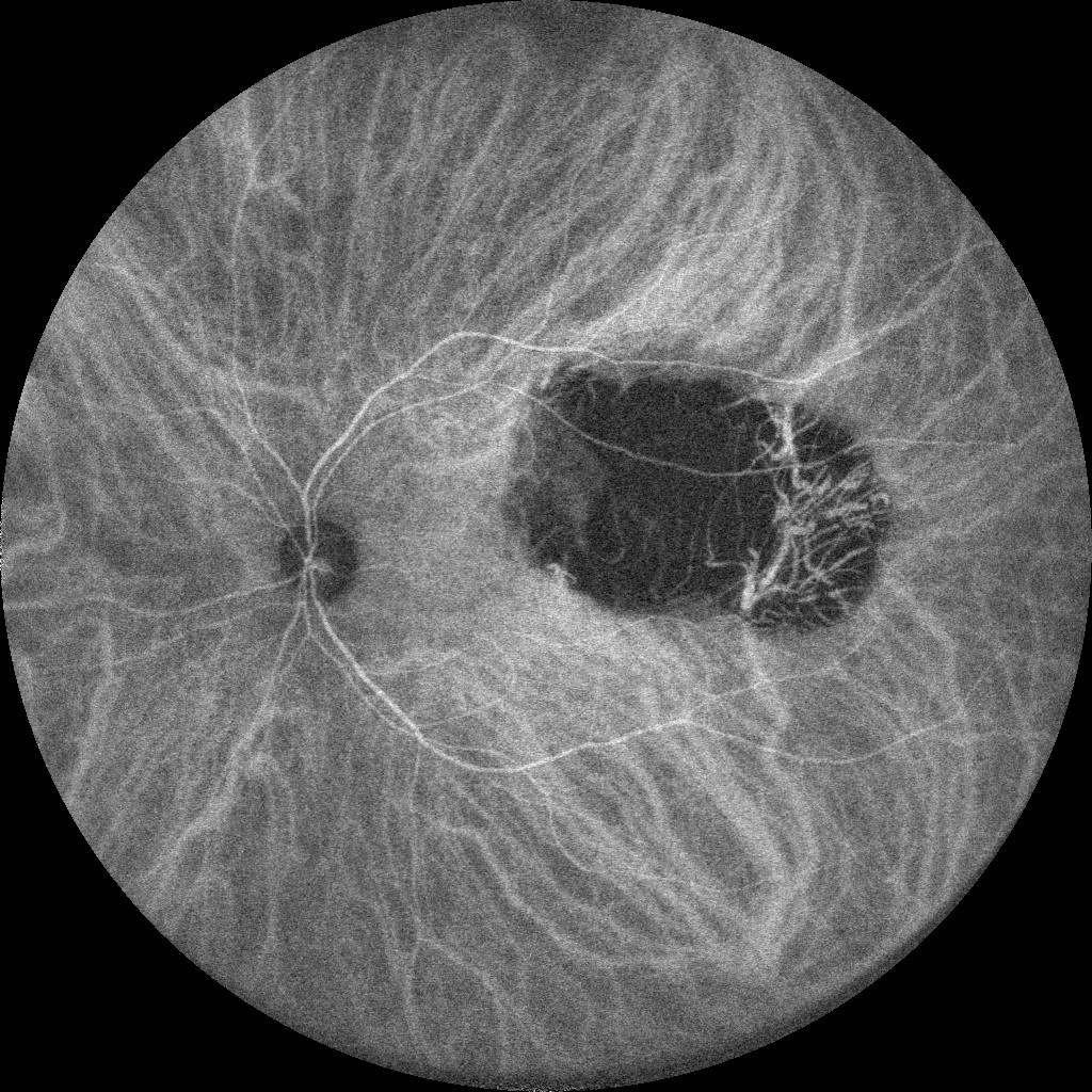
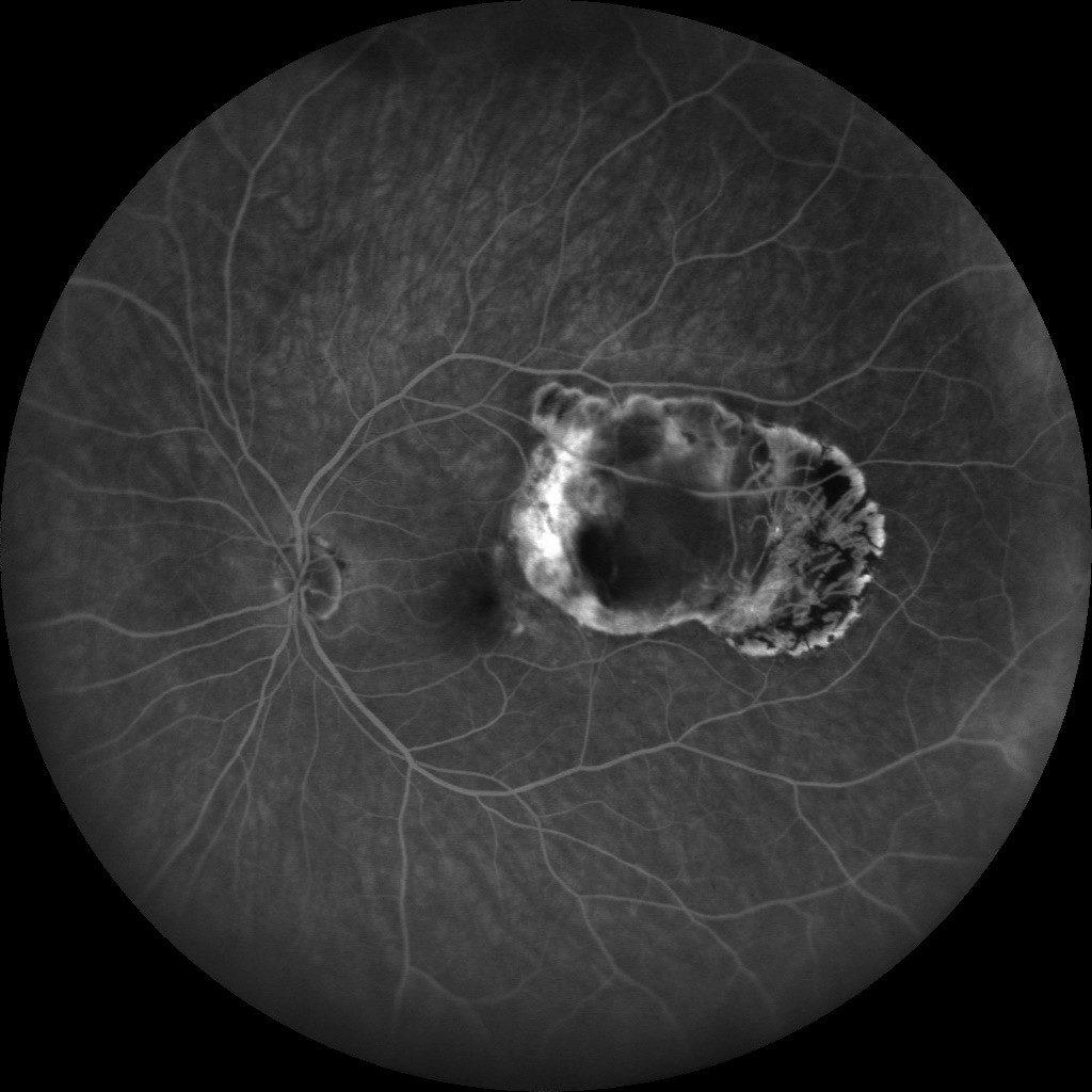
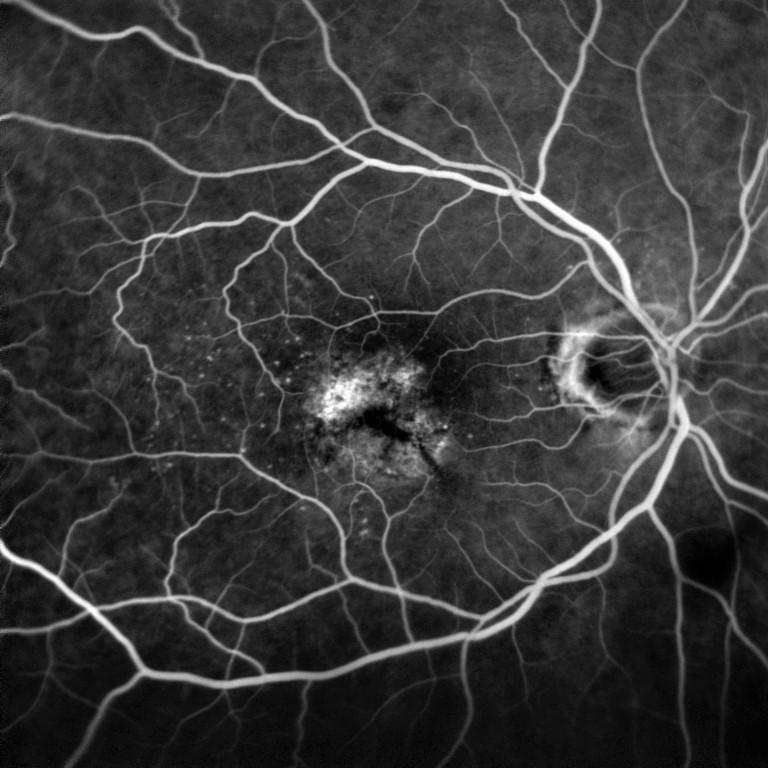
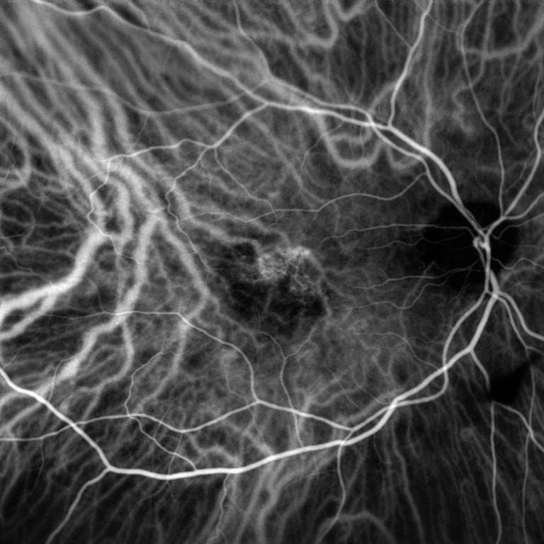
Pathologies that can be detected using an angiograph
Image courtesy: Dr Djaborouti (Puilboreau, France), hospital Luigi Sacco (University of Milan, Italy), hospital Lariboisière (Paris, France).
The different types of angiography
Benefits of angiography
The angiography is the only technique realising a dynamic observation of fundus vascularisation. Depending on the lens used, the analysis zone can be extended to make possible detecting some early vascular anomalies.

- Dynamic analysis
Dynamic analysis of retinal and choroidal vascularisation

- Extended acquisition
Visualisation du fond d’œil jusqu’à l’extrême périphérie grâce au grand champ

- Assistance to diagnosis
The vascular analysis completes the structural analysis performed using fundus photography and the OCT to confirm and refine the diagnosis.
Do you want to purchase an angiograph?
You have a project? You want a quotation? You have questions about our products? Feel free to ask your technical sales representative.
The Art of Eye Care
Reliability and
safety
NIDEK develops its top-of-the-range products to improve visual health through an approach based on strict criteria: safety, reliability, durability, continuous quality controls and certifications.
Technologies and
innovations
NIDEK meets technical challenges by keeping constantly informed of the innovations of eye imaging systems, using the expertise of professionals and the progresses of research.
Services and
guarantees
NIDEK commits itself to providing services to its customers, from the installation of an activity to the authorised training of teams, and to offering long-time measurable guarantees.
L'actualité NIDEK
🎉 NIDEK à la SFO 2025 : notre gamme médicale à l’honneur !
🎉 NIDEK à la SFO 2025 : notre gamme médicale à l’honneur ! Du 10
NIDEK accompagne la formation des assistants médicaux en ophtalmologie avec Keyce Santé
Chez NIDEK, nous sommes convaincus que l’innovation ne se limite pas aux équipements : elle
Retour sur la visite de M. Ozawa, président de NIDEK, en France
Le 11 février, NIDEK SA France a eu l’honneur d’accueillir M. Ozawa, président de NIDEK,
Index Égalité Femmes-Hommes 2024
Index Égalité Femmes-Hommes 2024 : Nidek SA progresse avec un score de 87/100 Chez Nidek
🎉 NIDEK à la SFO 2025 : notre gamme médicale à l’honneur !
🎉 NIDEK à la SFO 2025 : notre gamme médicale à l’honneur ! Du 10
Retour sur la visite de M. Ozawa, président de NIDEK, en France
Le 11 février, NIDEK SA France a eu l’honneur d’accueillir M. Ozawa, président de NIDEK,
Nidek et MEDADOM renouvellent leur partenariat pour leur dispositif innovant de téléophtalmologie
Communiqué de presse Nidek et MEDADOM renouvellent leur partenariat pour leur dispositif innovant de téléophtalmologie
NIDEK SA à la Journée Nationale de Téléophtalmologie 2024
Nous sommes ravis d’annoncer la participation de NIDEK SA à la Journée Nationale de Téléophtalmologie

