Homepage > Multimodal platforms > Mirante (SLO/OCT)
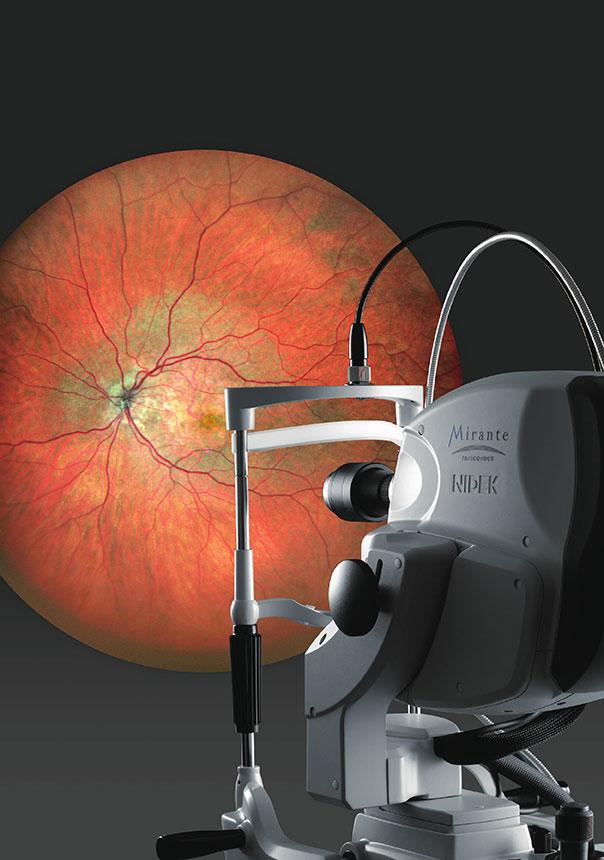
The Mirante is a large-field imaging multimodal platform designed to fundus imaging. It combines OCT and LSO imaging, thus grouping all routine imaging to detect and identify retinal-choroidal pathologies. By adding a lens, performing a section analysis of the anterior segment is possible.
The Mirante embraces in a single product all the imaging modalities used daily to observe the fundus. They can also be obtained in ultra large field mode:
It integrates a LSO (Laser Scanning Ophtalmoscope) that uses infrareds and provides high-contrast and high-resolution images. This systems also performs the tracking of the patient’s eye before and during the OCT mode acquisition, thus ensuring the correct positioning of OCT and OCT-Angiography scans by considering the natural cyclotorsion of human eye.
The images of the fundus obtained through an LSO scanning and a normal 89° field, and a large field up to 163° are high-definition images, with a premium contrast.
The analysis of colour photos and red, green and blue original images, obtained directly and without a filter from each wavelength-dedicated sensor makes possible viewing the finest details of the fundus to easily detect the lesions arising in retinal-choroidal pathologies.
We use the auto fluorescence to analyse the pigmentary epythelium. There are two versions of auto fluorescence: the green wavelength one to correctly view the macula and the blue wavelength one, more convenient to observe the papilla. Therefore, adapting the reading to the studied pathology is possible.
Dynamic or static injection angiography acquisitions, using fluorescein and/or the ICG, are used to observe the vascularisation of the retina and the choroid, from the early filling phases to the late times. Thanks to the real-time infrared imaging, the alignment is performed at the beginning of the injection, thus reducing the loss of information. The automatic setting of the gain, independent for the fluorescein and the ICG, limits the saturation of the signal that sometimes hind the fundus. You can then confirm your diagnosis using the detection of vascular lesions, such as aneurysms, ischemia and leaks.
The retro mode is a technology specially attached to the Mirante. It is an infrared imaging of the deep layers of retina to highlight the structures of the pigmentary epythelium and of the choroid. It uses, derivatively, the LSO technology, and reinforces the detection of fine lesions on these difficult-to-access layers, particularly the lesions of drusen, the conventional imaging methods cannot sometimes see.
The ultra large field adapter is used to enlarge the analysis zone up to 163°, to access to the periphery. By tilting and rotating the upper part of the product, you can observe the fundus, up to the extreme periphery. The reconstruction of different pictures provides a view of the entire fundus, up to the ora serrata.
Designed to performed specialised consultations, the Mirante integrates different advanced OCT functions, such as the HD mode, the manual positioning of acquisition scans on the area of interest, the Select mode and Rescan, to perform an additional exam, or the following-up of patients, available as soon as the acquisition.
Thanks to the LSO Tracking, the Mirante can perform the following-up of examinations exactly in the same conditions, through the recognition of the patient’s retina. This ensures a reliable comparative analysis, including on a single section. Up to 50 examinations can be compared over time, to monitor the evolution of the disease and/or to control the efficiency of the cure. Use trend curves and add a manual event, such as the date of the first use of the therapy, for instance, to perform an efficient and tailor-made following-up
The Mirante also integrates an anterior segment adapter to make sections of the cornea and of the filtration angles.
The AngioScan makes possible the analysis of the retinal and choroidal vascularisation. The real time tracking of the retina ensures a correct positioning of the scans, to limit any motion artefacts. To make the analyses different, a picture is taken either of the macula (Macula Map), or of the optical nerve head (Disc Map). The segmentations are adapted to the captured zone, 7 different zones on the macula and 4 zones on the papilla, and the density and vascular perfusion values are given to perform an additional quantitative analysis.
Thanks to the combined OCT En-Face and OCT-Angiography display, whose depth can be adjusted, and to the B-scan section (from a drop-down menu), a direct matching can be made between the structure and the tissue vascularisation.
The NAVIS-EX software platform can analyse all the data. This interface controls the entire patient database for the NIDEK imaging devices. It is equipped with viewers dedicated to Mirante results.
LSO imaging analyses of the fundus:
OCT and OCT-Angiography analyses
These mentions are conforms to the French regulation and may vary depending on circumstances in each country.
Brochure et livrets de cas cliniques
Articles multimodalité :
Retours d’expert
Retours d’expert rétromode :
Présentation de cas
https://retinatoday.com/issues/2022-may-june-insert3
You have a project? You want a quotation? You have questions about our products?
Feel free to ask your technical sales representative.
Reliability and
safety
Technologies and
innovations
Services and
guarantees
KEY PRODUCTS
© 2021 • NIDEK SA – All rights reserved
Ce site est réservé aux professionnels de santé