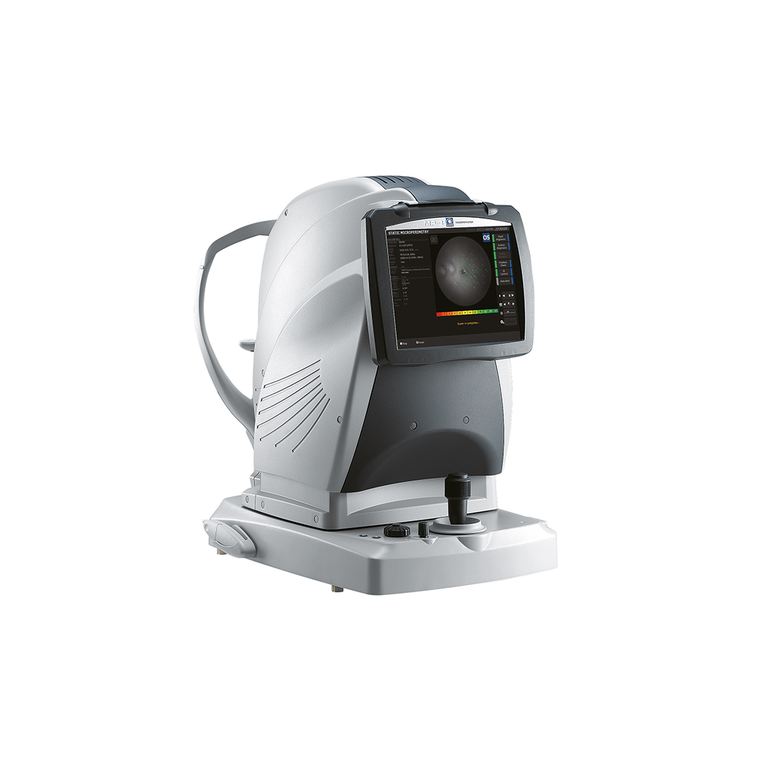
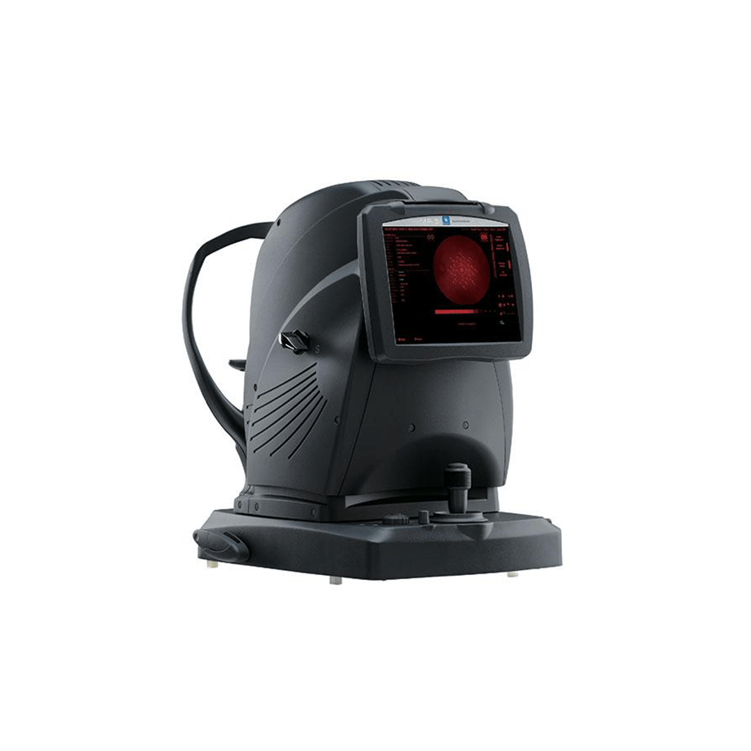
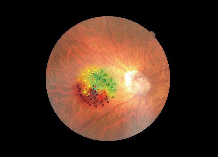
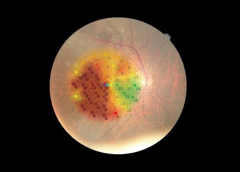
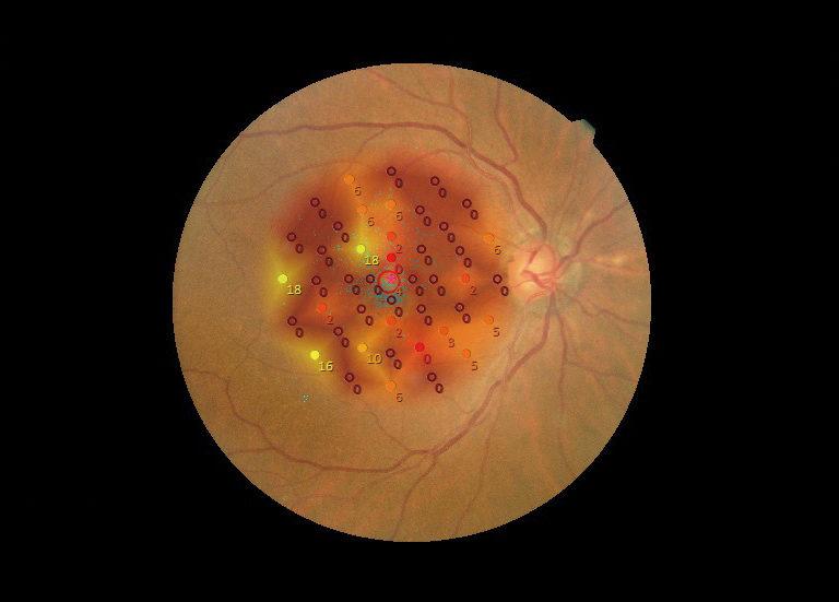
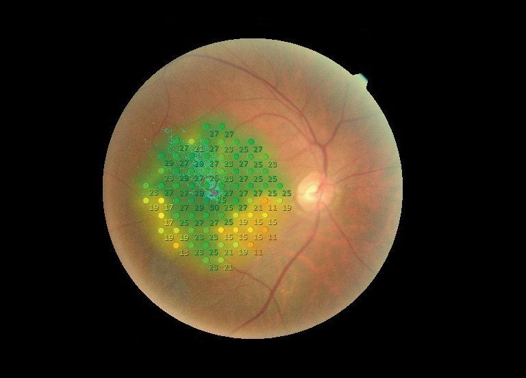
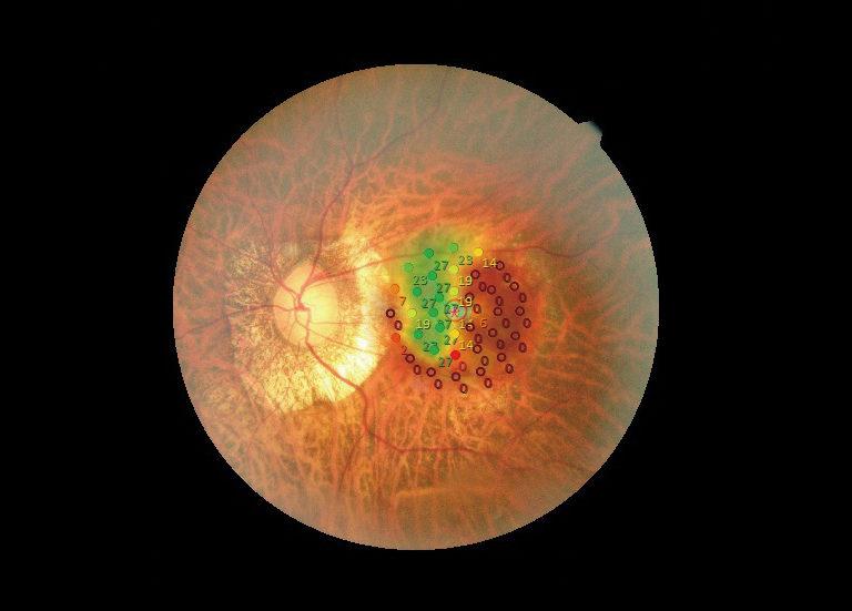
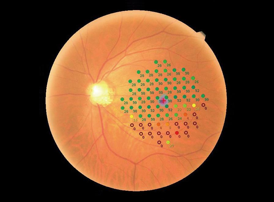
The purpose of microperimetry is assessing the retinal sensibility, the running of the photoreceptor cells of the eye, of the cones and rods, using light stimuli proposed to the patient. The stimuli are emitted with different intensities, with increasing or decreasing values (depending on the strategy selected to determine the threshold) in order to define the response thresholds of retina. Each time the patient detects the stimulus, he/she presses the response bulb, thus indicating to the instrument that he/she has got the intensity of this stimulus. Once the stimulus is not detected any more, the maximal sensibility is set (in dB).
Unlike conventional visual field devices, microperimeters have a three-dimensional retinal tracking system for accurate positioning of stimuli on a selected zone of the retina and therefore the analysis area.
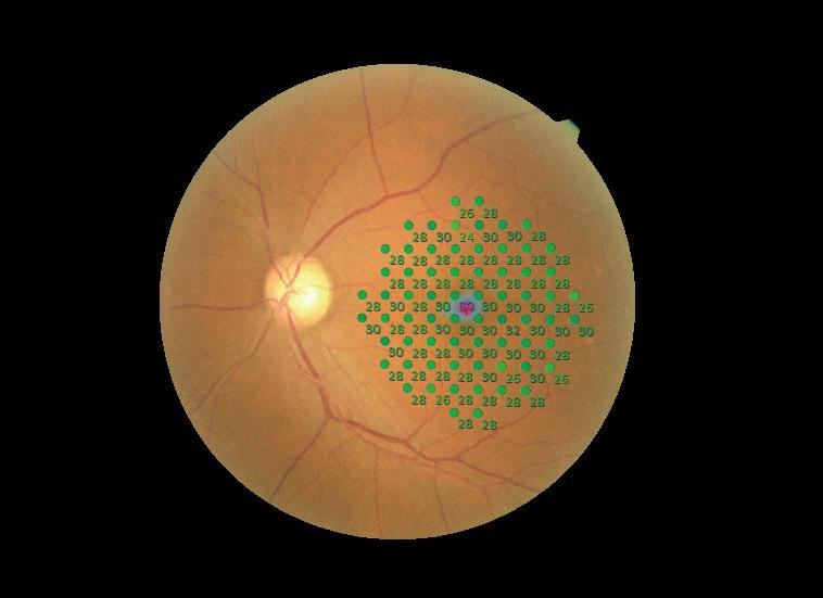
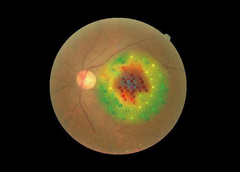
The results are presented on a fundus, in order to view the position of the stimuli and their threshold value. Also, thanks to tracking, the microperimeter follows the patient’s fixation during the microperimetry examination to define fixation stability and evaluate overall visual quality. This information is then used to define visual rehabilitation strategies, primarily in patients with very low visual acuity (low vision).

