Homepage > Microperimeters > MP-3
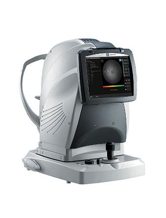
The MP-3 is used to perform the microperimetry exam. It is a very accurate examination used to perform the functional analysis of the retina photoreceptor cells, of the cones and rods, in both photopic and mesopic media. This examination comes with a colour photography of the fundus on which the results superimpose to improve the understanding.
The MP-3 is a microperimeter equipped with a 3D tracking system of the eye and a non-mydriatic fundus camera used to perform microperimetry, to assess the sensibility of photoreceptor cells of the retina. The range of light intensities, from 0 to 34 dB and the max. luminance of stimuli at 10,000 abs makes possible detecting the lowest sensibilities.
This device is equipped with a 3D tracking system of the eye, real time, to correctly position the light stimuli on the retina and limit the patient’s fatigue, as it is quicker. During the examination, the stability of the fixation is also analysed to complete the diagnosis and follow-up the progress of the patient’s rehabilitation. This examination can be performed on its own. It lasts 40 seconds. The analyses are presented either on 2° and 4° circles or in BCEA (Bivariate Contour Ellipse Area).
The device itself, thanks to the marks given by the fundus, tracks and aligns to the patient’s eye (anterior and posterior segments) and positions again the results to perform a correct matching with the photography of the fundus. This technology is also used to quantify the evolution of the patients’ sensibility by comparing the similar test zones through a following-up. Then the second examination is automatically carried out in the same conditions as the first one.
The active rehabilitation of the patient (feedback function), mainly used for low-vision patients (with a very low visual acuity), is easily performed, in an optimised way, thanks to the sound guidance (beeps) and the additional visual stimulation (flickering) This procedure reduces the duration of rehabilitation sessions (10 min instead of 30-40 min).
The NAVIS-EX software interface provides an intuitive display of the results and proposes to the user the ability to totally set the examination, particularly using a 100%-adjustable stimuli grid. Thanks to this software, superimposing easily the microperimetry results to the patient OCT exams is possible, to perform a deeper multimodal analysis. To compare the results obtained with the classical visual field devices, a similar Humphrey gray scale is also available.
These mentions are conforms to the French regulation and may vary depending on circumstances in each country.
Retours d’expert :
Articles :
You have a project? You want a quotation? You have questions about our products?
Feel free to ask your technical sales representative.
Reliability and
safety
Technologies and
innovations
Services and
guarantees
KEY PRODUCTS
© 2021 • NIDEK SA – All rights reserved
Galerie
Ce site est réservé aux professionnels de santé