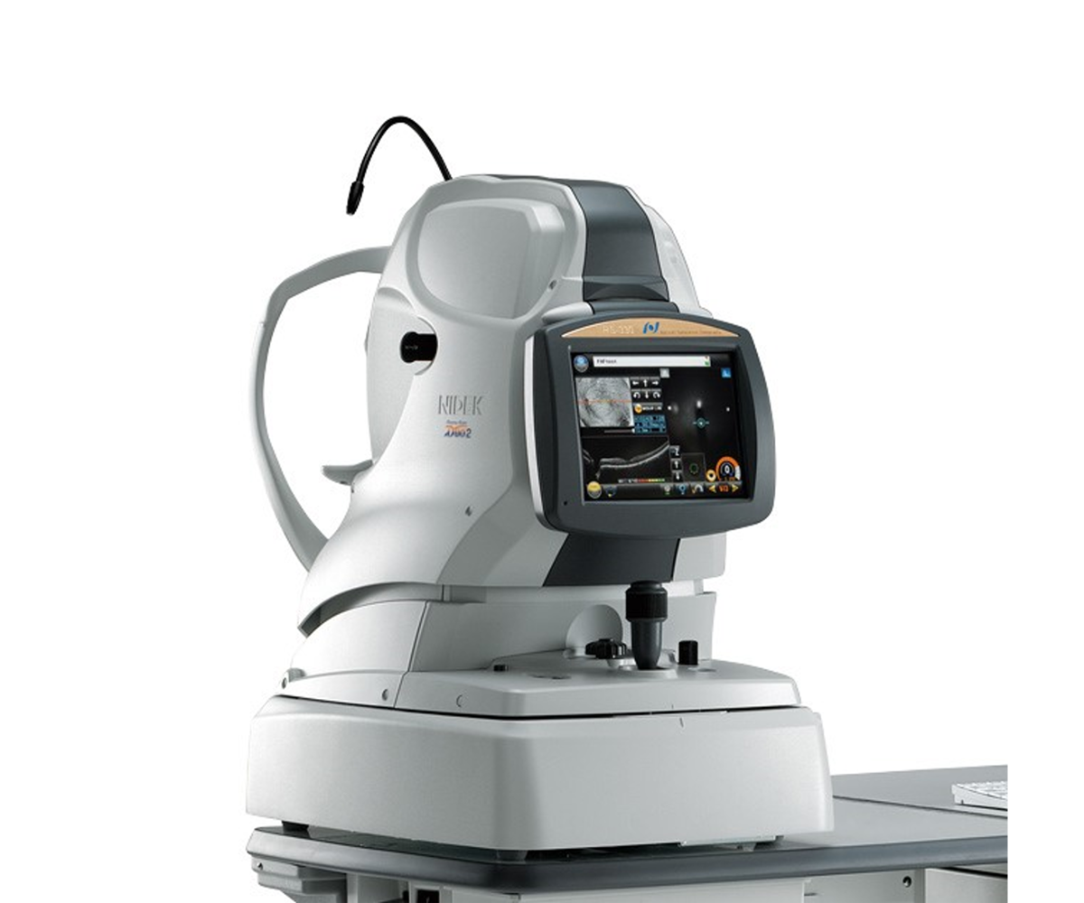
The Retina Scan Duo 2, combined fundus and autofluorescence OCT, is equipped with a 3D eye tracking system and automation to simplify measurement.

The Retina Scan Duo 2, combined fundus and autofluorescence OCT, is equipped with a 3D eye tracking system and automation to simplify measurement.
For ease of use, the device tracks the patient’s eye in real time and performs pupil alignment, retinal focus and image capture (OCT + photo). With its new Retina Map scan, combining macular and papillary analysis in a single capture, the Retina Scan Duo 2 is perfect for routine use in screening and standardising examinations. Signal sensitivity (Regular, Fine or Ultra-Fine mode) can be modified to increase signal intensity in the event of media opacity.
Depending on the configuration of the consultation, you can install the device on a NIDEK dedicated lifting table (depending on the model) for a daily use.
Acquisition fields of up to 12×9 mm in OCT and 45° in retina photography and autofluorescence offer multimodal analysis of the fundus and allow early detection of certain macular pathologies thanks to autofluorescence in the green wavelength, which is absorbed less by the macular pigment.
For greater expertise, other imaging modalities are also possible, several OCT slices are available as well as an average of up to 50 slices (HD mode), EDI mode for visualisation of the choroid, and photocolor and OCT modes for the anterior segment. Optionally, structural analysis can be supplemented by observation of retinal-choroidal microvascularisation using the AngioScan module.
Once acquired, the data is displayed on the NAVIS-EX software platform equipped with a OCT result and photograph viewer. Thickness maps, total retinal thickness and complex thickness of the ganglion cells are displayed and compared with the normative database in 9x9 mm, and nerve fibres of the optic nerve head are presented in 6x6 mm.
The software also provides En-Face OCT representation, as well as colour photos on which measurements such as papillary Disc and Cup can be taken.
AngioScan allows analysis of retinal and choroid vascularisation. The image is taken either of the macula (Macula Map) or of the head of the optic nerve (Disc Map), thus differentiating analyses. Segmentations are adapted to the scanned area, with seven differentiated areas on the macula and four on the papilla.
Thanks to the combined OCT En-Face and OCT-Angiography display, whose depth can be adjusted, and to the B-scan section (from a drop-down menu), a direct matching can be made between the structure and the tissue vascularisation.
The results of the AngioScan also include quantitative analyses. These additional parameters are used to make a diagnosis. Density and vascular perfusion values are given for each layer, as well as the information related to the central avascular zone (CAZ), which is automatically detected.
Brochures
Articles
You have a project? You want a quotation? You have questions about our products?
Feel free to ask your technical sales representative.
Reliability and
safety
Technologies and
innovations
Services and
guarantees
KEY PRODUCTS
© 2021 • NIDEK SA – All rights reserved
Ce site est réservé aux professionnels de santé